Tilt series/Tomography
Three-dimensional information of biological or soft materials samples including their surrounding environment can be obtained by collecting a Cryo Tilt Series. Integrity and preservation of the structure of vitrified specimen from beam damage must be ensured, as the sample is typically exposed to higher total dose than for Single Particle techniques.
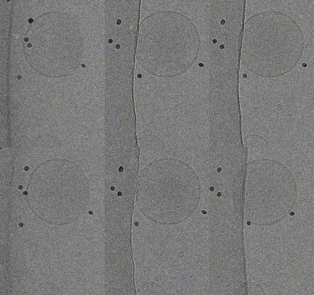
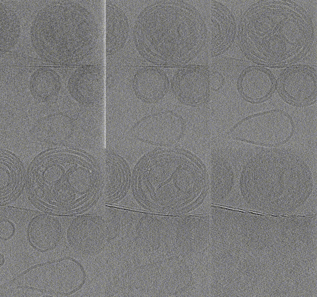
The target area is imaged under low-dose conditions at different angles by rotating the microscope stage. After alignment of the images at different angles, the tilt series can be inspected to gain 3D information about the sample (see below, left). Furthermore, the tilt series can be back projected to reconstruct a 3D volume i.e., obtain the cryo electron tomogram or cryo-ET (see below, right).
The cryoET technique can also be applied in cellular context and combined with template matching to gain molecular level information (numbers, locations) of large macromolecular machines such as ribosomes, proteasomes etc. in the cell. Moreover, sub-tomogram averaging techniques can be applied to further improve resolution of target macromolecule such as ribosome by averaging and aligning techniques in a similar to, but more complex fashion than Single Particle Analysis (SPA).


