In-Situ / Operando TEM
Selected projects performed at ScopeM using in-situ techniques are listed below.
Liquid Cell and Electrochemistry
Molecular catalyst design for water-splitting reactions
Water-splitting reactions as sources of molecular hydrogen are very promising routes for renewable energy storage solutions. The indispensable component of the controllable water-splitting processes, the oxygen-evolution reaction (OER), relies on catalytic efficiency of synthesized compounds that induce and promote the OER, for example the first-row transition-metal-based complexes.
The molecular mechanism of water oxidation mediated by metal complexes is however very complex and not fully understood. Transmission electron microscopy group of ScopeM in collaboration with the group of Professor external page Greta R. Patzke performs the dynamic studies of the transition metal mediated OERs by the in-situ electro-chemical liquid phase TEM (EC-LPTEM) experiments using GrandARM “Vortex” instruments and external page Protochips “Poseidon” liquid cell set-up.
The studies on the Cu- and Co-complexes based mediators indicate that the true catalysis of the OERs are the Cu- and Co-oxide nanoparticles rather than the molecular Cu- and Co-compounds as it was considered. The studies show that careful analytical approach and in-situ protocols are the necessity for the catalysts development.
The movie-example presents the results of the in-situ EC-LPTEM external page study by external page Esmael Balaghi and Alla Sologubenko on Cu-based complexes mediated OER and shows the formation of the Cu-O nanoparticles, the true catalytic entities of the OER.
Heating
Ferroelectric domains in multiferroic materials
Studying the ferroelectric domains, domain walls and phase transition in ferroelectric materials has become an active field of research in electron microscopy. The Materials Theory group in the Department of Materials, ETH, led by Prof. Nicola Spaldin, develops novel multiferroic materials and in collaboration with ScopeM, the ferroelectric domains, chemistry, and defects in multiferroic oxides on the nanoscale are investigated.
The sub-Å resolution of the aberration-corrected JEOL GrandARM electron microscope at ScopeM allows researchers to investigate nanoscale phenomena in ferroelectric materials. The images show multiferroic samples.
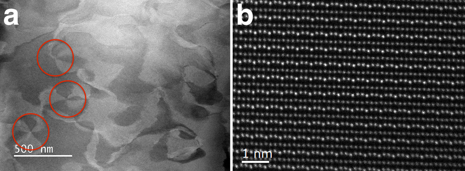
- Dark field TEM image of vortex ferroelectric domains in doped hexagonal RMnO3 oxides; see red circles in image (a) showing six-fold vortices
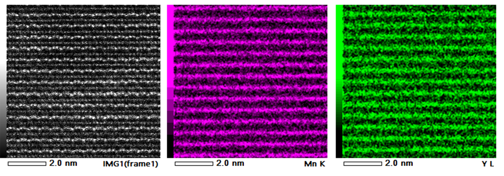
- HAADF-STEM image of YMnO3 single crystal; the HAADF detector is used to look at the atomic fluctuation of rare-earth ions; this usually requires better than 100 pm resolution, like in image (b)
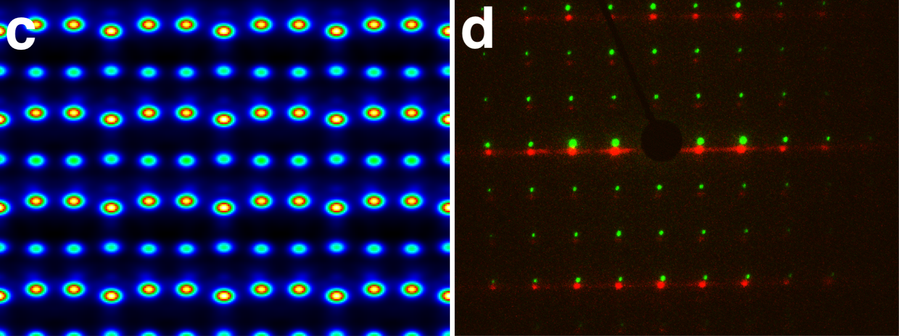
- STEM image simulation of RMnO3 lattice along [110] zone axis, shown in image (c), is used to better interpret images (Dr. Probe software package)
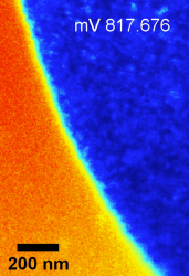
- Diffraction patterns of YMnO3 lamella at 100 °C and 700 °C along [110] zone axis using Protochip heating holder, as in image (d), is used to study phase transition
- The elemental (EDS) analysis of the lattices in h-RMnO3 oxides with lattice or atomic resolution is an additional tool to help to investigate the chemistry of the sample (image below)